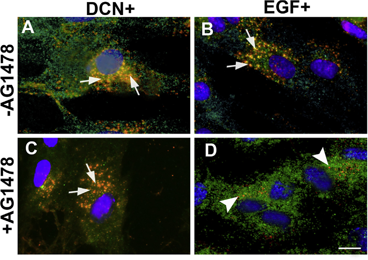
Fig. 5. Decorin-induced internalization of EGFR does not require tyrosine kinase activity.

Confocal microscopy of hCSF cells pretreated with AG1478 (a specific tyrosine kinase activity inhibitor at 5 μM for 60 min) and incubated with rhDcn (250 nM) do not inhibit internalization of EGFR and colocalize with Dcn (yellow; arrows) in the perinuclear space (C) similar to the non-treated cells (A). On the contrary, rhEGF-treatment (100 ng/ml) internalizes of EGFR was inhibited by AG1478 and EGFR expression (D) was seen only at the cell surface (green, arrowheads) while non-treated cells show internalization of EGFR + EGF (yellow; arrows). Merged image shows co-localization of EGFR + EGF observed near peri-nuclear region (B). Bar = 25 μM. (For interpretation of the references to colour in this figure legend, the reader is referred to the Web version of this article.)
