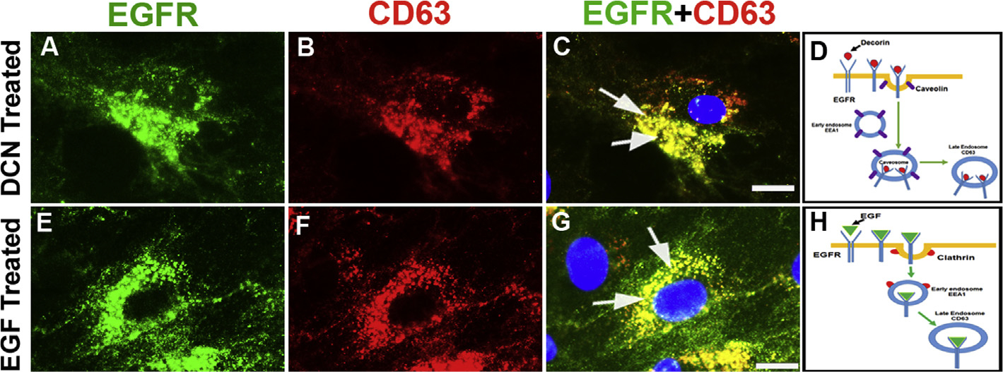
Fig. 7. Decorin-EGFR complex degradation involves late endosome.

Human CSFs incubated with rhDcn (A–D) or rhEGF (E–H) and double-immunostained with EGFR (green) and late endosome marker, CD63, (red). Decorin treatment showed high co-localization of EGFR and CD63 in an overlay image (C; yellow, arrows) compared to the rhEGF (G, yellow, arrows). The EGFR staining (green) is shown in panels A and E, and CD63 staining (red) is shown in panels B and F. Schematic diagram shows EGFR internalization target late endosome upon decorin treatment (D) compared to EGF treatment (H). Bar = 10 μM. (For interpretation of the references to colour in this figure legend, the reader is referred to the Web version of this article.)
