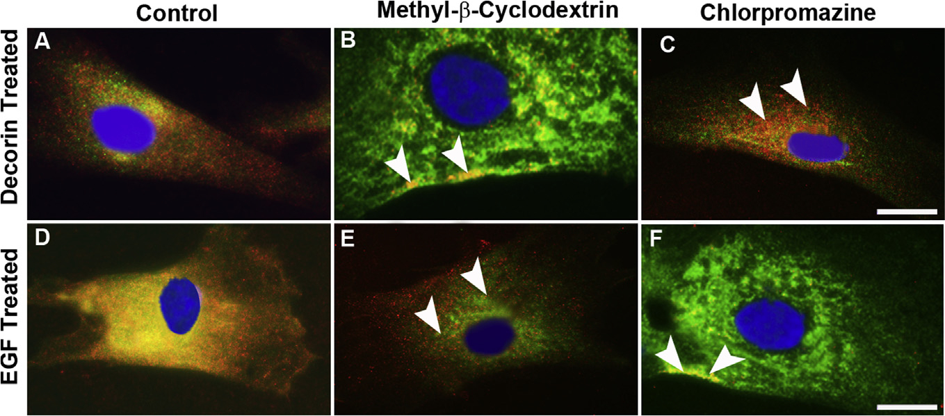
Fig. 9. Decorin-mediated internalization of EGFR ensues via caveolar-mediated endocytosis.

Confocal photomicrograph showing inhibition of caveolae formation in hCSF cells using methyl-β-cyclodextrin (MβCD) that depletes cholesterol from the plasma membrane, rhDcn-induced internalization of EGFR was completely blocked (B) and EGFR was localized only in the plasma membrane (arrowheads). Contrary to this, rhEGF-induced internalization of EGFR was not affected (E, arrowheads) when treated with MβCD. When clathrin assembly was inhibited by chlorpromazine, a cationic amphiphilic drug that preferentially blocks the receptor recycling by preventing the adapter protein AP-2 of clathrin assembly on clathrin-coated pits, rhDcn-induced internalization of EGFR was observed (C, arrowheads) while rhEGF-induced internalization of EGFR was disrupted (F, arrowheads). The vehicle-treated (-dcn) showed no EGFR internalization as shown in Fig. 4A. Bar = 5 μM.
