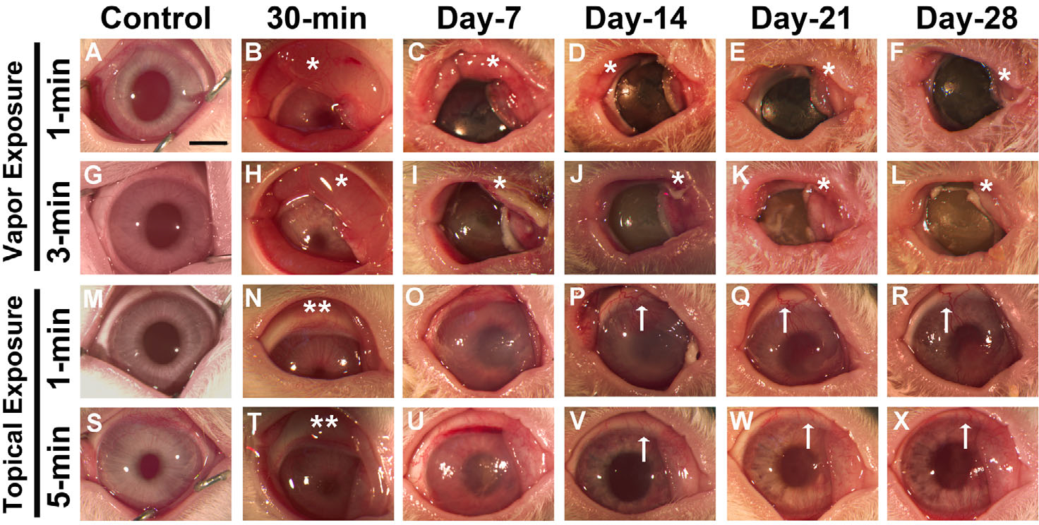
Figure 2.

In vivo stereobiomicroscopy images showing severe ocular damage in rabbit eyes from 30 min to 28 days following acrolein vapor exposure for 1 min (B–F) and 3 min (H–L) and topical exposure for 1 min (N–R) and 5 min (T–X). Panels A, G, M, and S are images of control nonexposed rabbit eyes. Vapor exposure to the cornea caused severe eyelid inflammation (*) and corneal opacity. Abscesses appeared in the conjunctival region, and there was severe corneal haze and an opaque corneal surface. Topical exposure to the eyes caused mild eyelid inflammation (**) in the early phase and prominent neovascularization (↑) in the late stages (14–28 days) of corneal wound healing. n = 6 rabbit eyes in each group. All images were taken at 7.1× magnification. Scale bar = 3.0 mm.
