Figure 3.

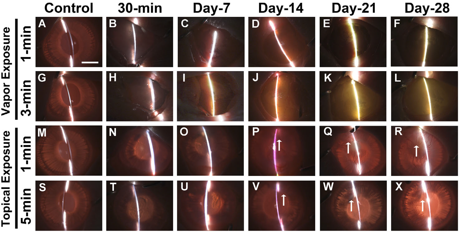
In vivo slit-lamp biomicroscopy images showed severe ocular opacity in the rabbit eyes from 30 min to 28 days after acrolein vapor exposure for 1 min (B–F) and 3 min (H–L) and topical exposure for 1 min (N–R) and 5 min (T–X). Panels A, G, M, and S are images of control, nonexposed rabbit eyes. Vapor exposure to the cornea caused thick bands and bright reflective areas, indicating severe corneal opacity. A similar pattern was observed after topical exposure, with growth of new vessels (↑) 21 and 28 days after acrolein treatment. n = 6 rabbits in each group. All images were taken at 7.1× magnification. Scale bar = 3.0 mm.
