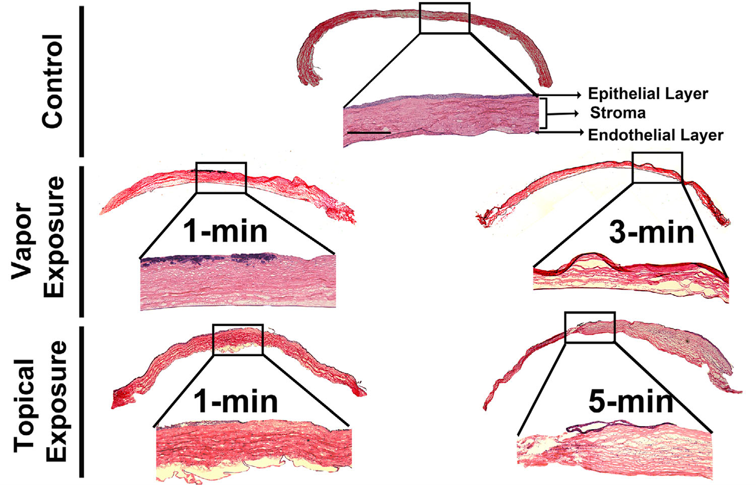
Figure 6.

Hematoxylin and eosin staining of the corneal sections of the control and acrolein-exposed rabbit eyes at day 28. Vapor-treated corneas showed loss of epithelial cells in the anterior stroma in the 1-min exposed group. On the other hand, the 3-min exposed group caused severe disruption of the corneal stroma as well as epithelial cell loss. One-minute topical exposure caused corneal edema and loss of epithelial cells as well as distorted cellular architecture with increased pathology in the 5-min exposed group, accompanied by infiltrating inflammatory cells in the corneal stroma. n = 6 rabbit eyes in each group. The entire corneal tissue section images are a montage of the 50× magnification and inset images. Scale bar = 100 μm. All inset images were taken at 100× magnification.
