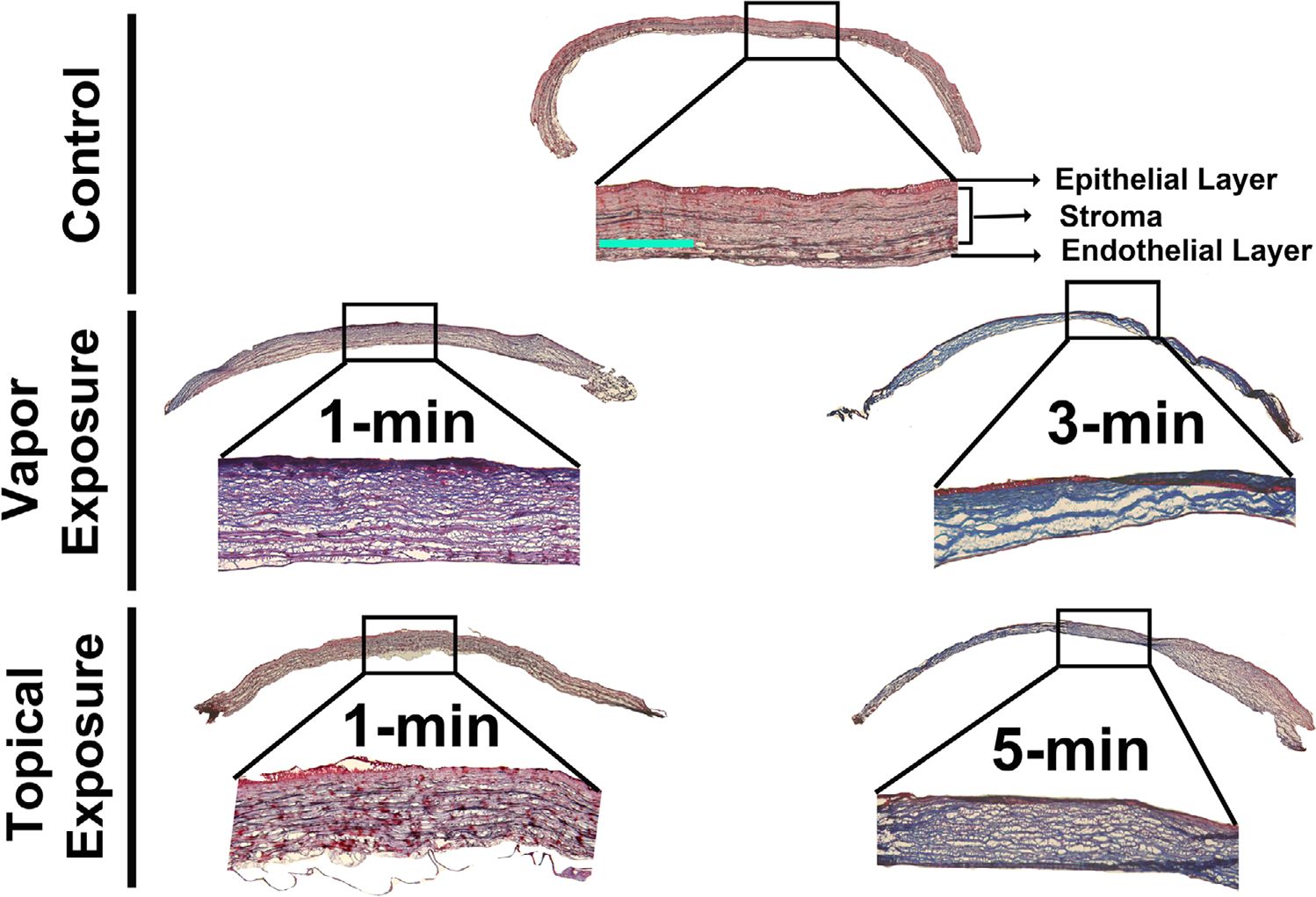
Figure 7.

Masson’s trichrome staining of the corneal sections of the control and acrolein-exposed rabbit eyes for the evaluation of corneal stromal collagen at day 28 after acrolein exposure. Vapor-treated corneas showed irregular collagen staining (collagen is stained blue) in the 1-min exposed group, which increased in staining intensity in the 3-min exposed group. The corneal collagens are highly disorganized and missing in certain areas due to the loss of corneal architecture. The topical exposure group showed similar collagen damage in the 1-min exposed cornea, with additional edema, and the staining intensity increased further in the 5-min exposed group, showing further damage and irregularity in the corneal collagens after acrolein exposure. n = 6 rabbit eyes in each group. The entire corneal tissue section images are a montage of the 50× magnification and inset images. Scale bar = 100 μm. All inset images were taken at 100× magnification.
