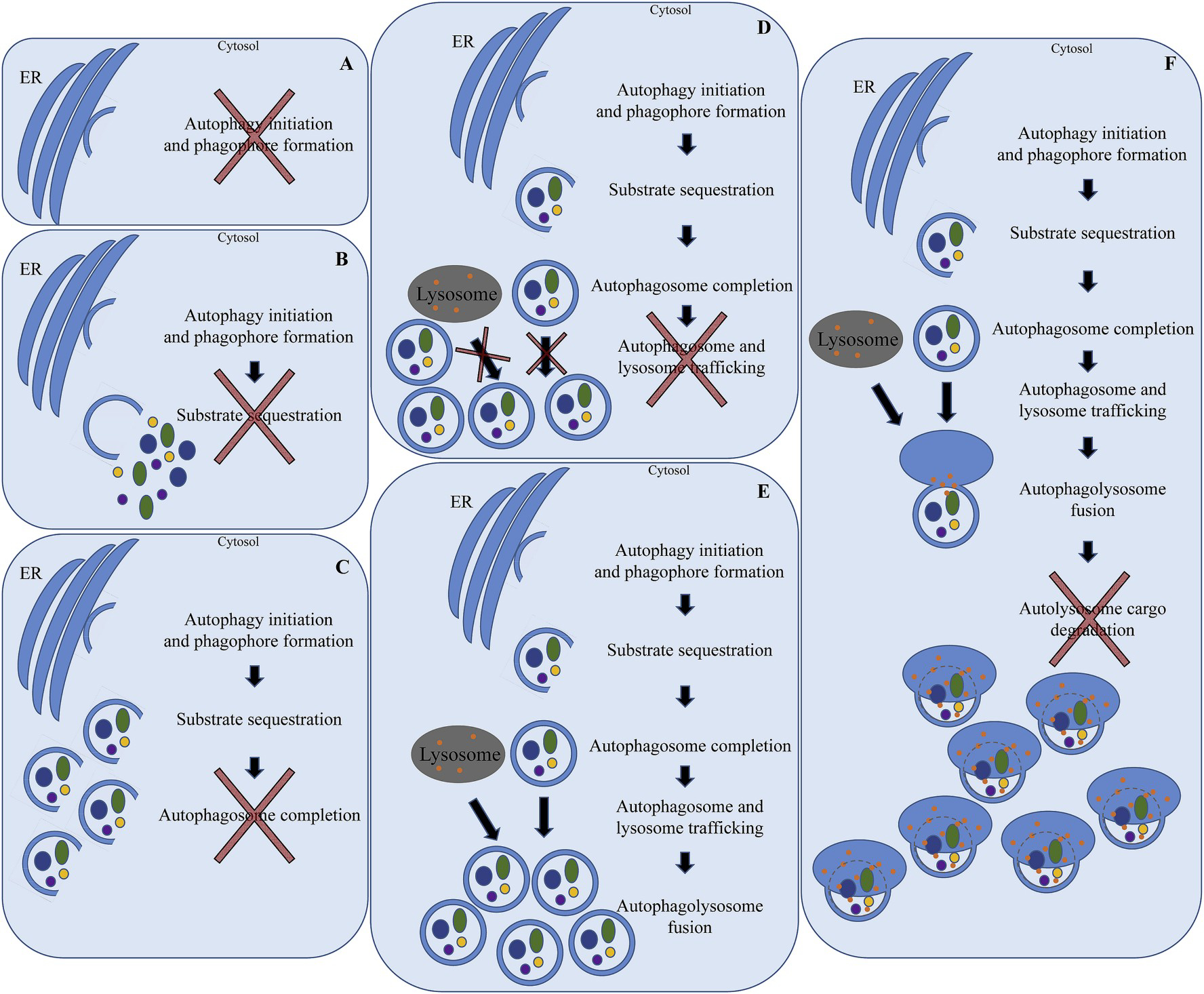
Fig. 4. Schematic of normal autophagy function (center) and the various points of failure that might result in autophagy dysfunction.

A) Failure of initiation cell signaling pathways to start phagofore formation. This might also occur due to lack of substrate from the endoplasmic reticulum. B) Failure of sequestration of cargo into the forming autophagosome. This might occur due to failed targeting of selected substrates or failed signaling and trafficking of selected substrates. C) Failure of complete double-membrane synthesis (elongation) of the developing autophagosome. This might result in either accumulation of partially formed autophagosomes or failed synthesis of the initial forming autophagosome. D) Failure of lysosome and autophagosome vesicular trafficking, which results in accumulation of autophagosomes. E) Failure to fuse the lysosome and autophagosome, which results in accumulation of autophagosomes. F) Failure to degrade autolysosomal sequestered cargo, which results in accumulation of autolysosomes.
(ER: endoplasmic reticulum).
