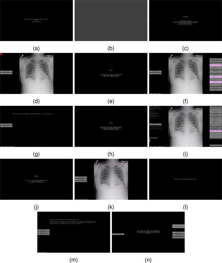
Fig. 2.
Screens of the data-collection interface in the sequence they are presented to a radiologist, including instruction screens (a,c,e,h,j,n), calibration of pupil size (b), dictation of reports (d), choice of global labels (f,g), selection of ellipses and certainties (i), drawing of lung/heart box (k), and editing of transcription (l,m). Digital visualization is recommended for reading the content.

