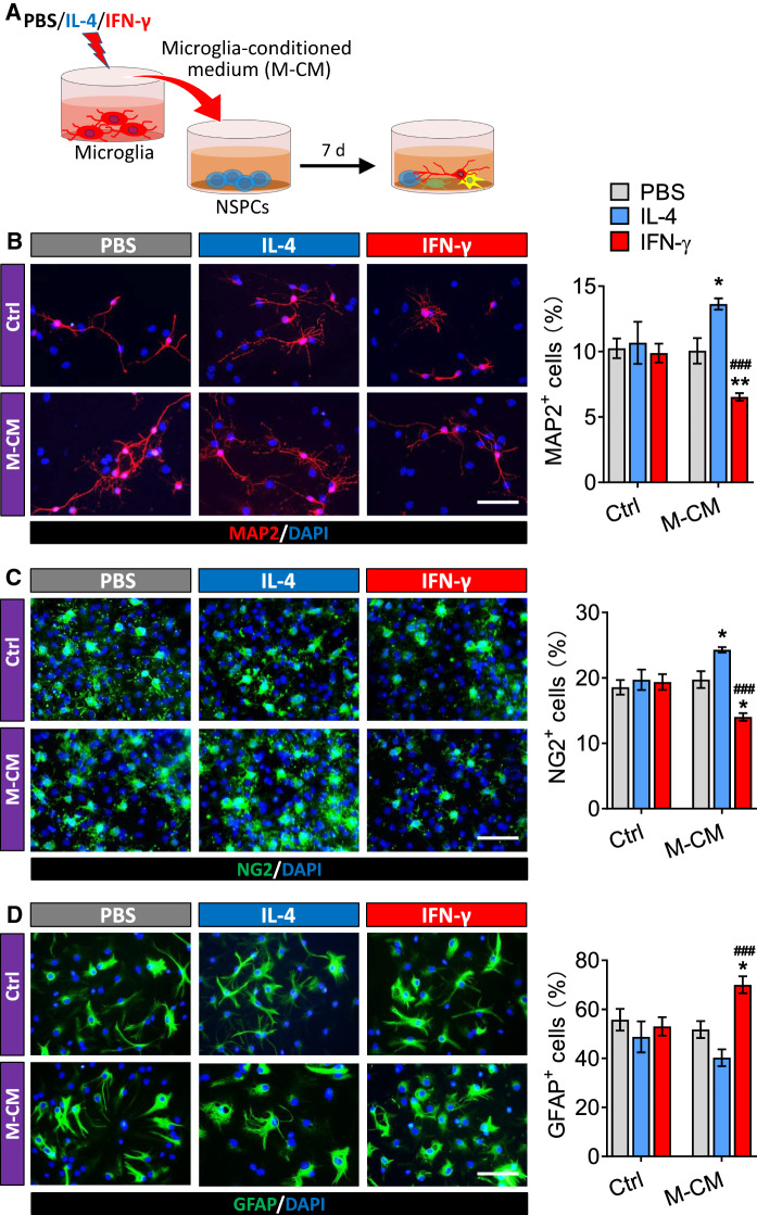
Fig. 6.
Effects of the secretome from IL-4- or IFN-γ-treated microglia on differentiation of NSPCs. A Experimental scheme to monitor microglia-conditioned medium on differentiation of adult NSPCs. B Fluorescence micrographs of MAP2+ cells from NSPCs differentiate in PBS, IL-4 or IFN-γ or microglia-conditioned medium for 7 days. The MAP2+ cells were stained with antibody (red) and nucleus is labeled by DAPI (blue). Scale bar is 10 μm. The histogram represents quantification of percentage of MAP2+ cells from adult NSPCs. C Fluorescence micrographs of NG2+ cells from NSPCs differentiate in PBS, IL-4 or IFN-γ or microglia-conditioned medium for 7 days. The NG2+ cells were stained with antibody (green) and nucleus is labeled by DAPI (blue). Scale bar is 10 μm. The histogram represents quantification of percentage of NG2+ cells from adult NSPCs. D Fluorescence micrographs of GFAP+ cells from NSPCs differentiate in PBS, IL-4 or IFN-γ or microglia-conditioned medium for 7 days. The GFAP+ cells were stained with antibody (green) and nucleus is labeled by DAPI (blue). Scale bar is 10 μm. The histogram represents quantification of percentage of GFAP+ cells from adult NSPCs. Data are showed Mean ± SEM, n = 4–6, *P < 0.05, **P < 0.01 vs PBS group, ###P < 0.001 vs IL-4 group

