Figure 2.

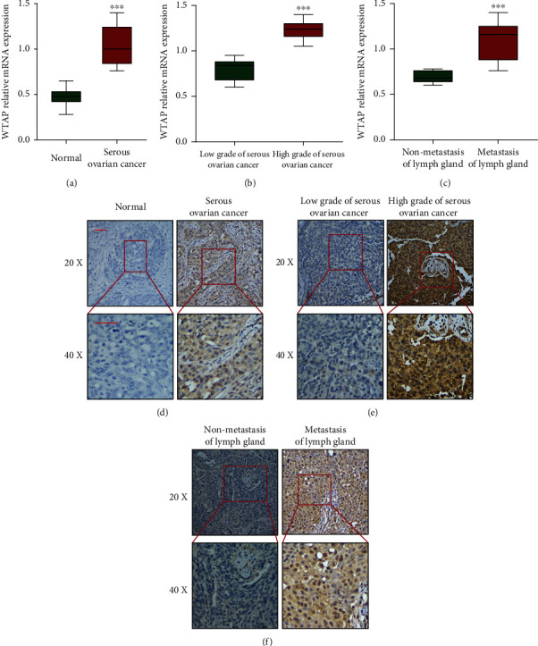
The expression of WTAP is significantly upregulated in ovarian cancer tissue and closely correlated with clinicopathological parameter. (a) The mRNA expression of WTAP was upregulated in serous OC tissues compared with normal tissues. (b and c) WTAP expression was relatively higher in high grade OC tissues and those with metastasis of the lymph gland. ∗∗∗P < 0.001. (d) Comparisons of WTAP expression in tissues revealed by IHC analysis in normal and serous OC tissues. (e) Comparisons of WTAP expression in tissues revealed by IHC analysis, low grade and high grade of serous OC tissues. (f) Comparisons of WTAP expression in tissues revealed by IHC analysis, nonmetastasis and metastasis of lymph gland tissues. The positive staining of WTAP was presented in brown color (distributed in both nucleus and cytoplasm), and the cell nuclei were counterstained with hematoxylin. Original magnification, 20x (left panels) and 40x (right panels). Scale bars = 10 μm.
