Fig. 1.
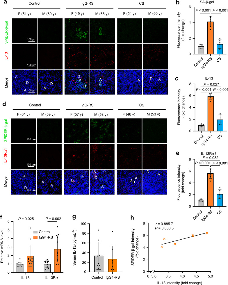
The number of senescent cells and expression of IL-13 and IL-13α1 are significantly increased in the SMGs of IgG4-RS patients. a–e Immunofluorescence staining (a, d) and quantitative analysis of SA-β-gal (b), IL-13 (c), and IL-13Rα1 (e) in the SMGs of IgG4-RS patients (n = 6), controls (n = 6), and CS patients (n = 6). A acinus, D duct, M male, F female, y years old. Bars = 100 μm. f Quantitative real-time PCR analysis showing the expressions of IL-13 and IL-13Rα1 in the SMGs of IgG4-RS patients (n = 12) and controls (n = 12). g IL-13 level in the serum of IgG4-RS patients (n = 8) and controls (n = 8) was detected by cytokine antibody array. h Correlation between the immunofluorescence staining intensity of IL-13 and the activity of SA-β-gal in the SMGs of IgG4-RS patients (n = 6) were analyzed by Spearman test. The significance of differences between groups was analyzed by unpaired Student’s t-test (f, g) or one-way ANOVA followed by Bonferroni’s test (b, c, e). Data are presented as the mean ± SD

