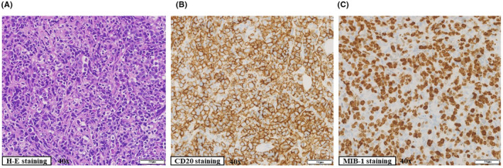
FIGURE 1.

Histopathological findings of right chest wall subcutaneous mass. Immunohistochemistry was performed on paraffin‐embedded tissue. (A) Diffuse proliferation of large lymphocytes. (B) Diffuse proliferation of CD20‐positive large lymphocytes. (C) The percentage of MIB‐1‐positive cells was more than 70%
