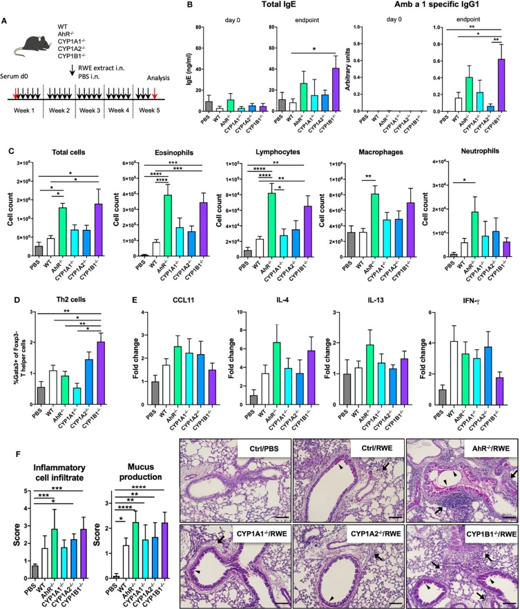
Figure 1.
AhR and CYP1B1 deficiency aggravates pollen-induced allergic airway inflammation. (A) Experimental setup. Mice of indicated genotypes were exposed for five weeks to RWE extract. Wildtype mice exposed to PBS served as negative controls (PBS). (B) Total IgE (left) and Amb a 1-specific IgG1 (right) at day 0 and endpoint. (C) Total and differential BAL cell counts, (D) Th2 cell frequency, (E) CCL11, IL-4, IL-13, IFN-γ expression in whole lung tissue and (F) histological scores (left) and representative lung sections (right). Arrows: inflammatory infiltrate; arrowheads: mucus hypersecretion; scale bar: 100µm. Mean ± SEM, n=6-21 mice/group from up to 3 independent experiments. Histological scores: mean ± SD, (n=5). ANOVA with Tukey’s multiple comparisons. *p < 0.05, **p < 0.01, ***p < 0.001, ****p < 0.0001.

