FIGURE 1.
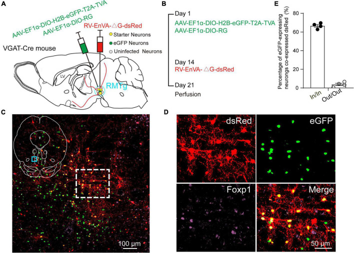
Virus injection and starter neurons in the rostromedial tegmental area (RMTg) in the vesicular GABA transporter (VGAT)-Cre mice. (A) Schematic diagram of injection of helper viruses, including adeno-associated virus (AAV) expressing tumor virus receptor A (TVA) with a green fluorescent protein (AAV-EF1α-DIO-H2B-eGFP-T2A-TVA) or expressing rabies glycoprotein (RG) (AAV-EF1α-DIO-RG) into the RMTg in a VGAT-Cre mouse, followed by injection of modified rabies virus (RV) expressing dsRed (RV-EnVA-△G-dsRed). (B) Experimental timeline for injection of helper viruses, RV, and perfusion. (C) Immunostaining showed the presence of merged neurons (yellow) in the RMTg. The dashed square region is the magnification of the indigo square region in the left upper islet. (D) Higher magnification of the dashed square region in C. Red, RV-infected neurons; green, helper virus-infected neurons with no RV infection; purple, neurons stained with Forkhead box protein 1 (Foxp1); yellow, starter neurons merged with both helper viruses and RV. (E) Co-expression rate of dsRed-labeled neurons in eGFP-expressing neurons. In, inside the RMTg; Out, outside the RMTg. n = 4, each data point represents one experimental mouse.

