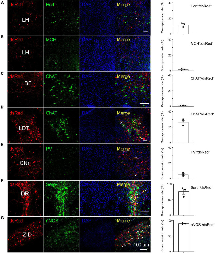
FIGURE 5.
Immunostaining of typical nuclei innervating rostromedial tegmental area γ-aminobutyric acid-releasing (GABAergic) neurons with several markers of sleep-wake regulation. (A–G) Images showing that dsRed-labeled neurons colocalized with hypocretin (Hcrt) and melanin-concentrating hormone (MCH) in the lateral hypothalamus (LH) (A,B), choline acetyltransferase (ChAT) in the basal forebrain (BF) and laterodorsal tegmental nucleus (LDT) (C,D), parvalbumin (PV) in the substantia nigra, reticular part (SNr), (E) serotonin (Sero) in the dorsal raphe nucleus (DR) (F), and neuronal nitric oxide synthase (nNOS) in the zona incerta, dorsal part (ZID) (G). The merged neurons are pointed by arrows. Rightmost column, quantification of dsRed-labeled cells that co-expressed with specific cell type biomarkers. n = 3, each data point represents one experimental mouse. Abbreviation: DAPI, 4’, 6-diamidino-2-phenylindole.

