
FIGURE 4.
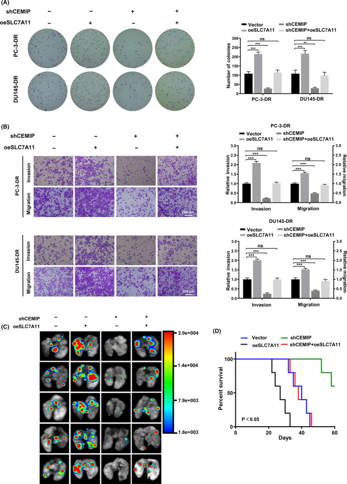
SLC7A11 rescues the invasion and metastasis of shCEMIP detachment‐resistant (DR) prostate cancer (PCa) cells in vitro and in vivo. (A) Representative images (left panel) and quantification (right panel) of soft agar plates indicating anchorage‐independent growth of the DR PCa cells stably transfected as indicated. (B) Transwell analysis of the invasion and migration capability of DR PCa cells stably transfected as indicated. Scale bars, 200 μm. (C) Representative image of metastatic lung colonization in nude mice (n = 5 per group) treated with tail vein injection of DR PC‐3 cells stably transfected as indicated. (D) Kaplan–Meier curves for nude mice. Data are presented as representative images or as mean ± SD from three independent repeats. **p < 0.01, ***p < 0.001