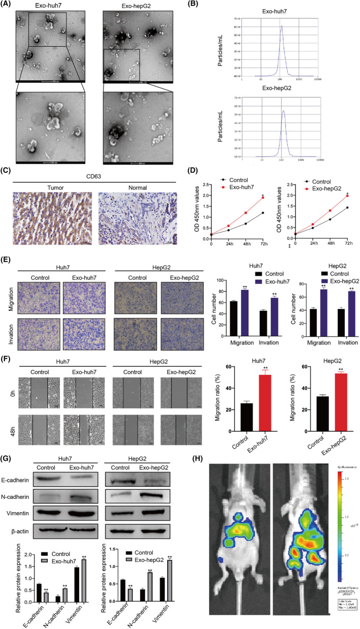
FIGURE 1.

Hepatocellular carcinoma (HCC)‐derived exosomes enhance HCC proliferation, migration, and invasion. A, Identifying the size and morphology of exosomes by TEM (scale bar = 200nm and 100nm). B, Identifying the size of HCC‐derived exosomes by nanoparticle‐tracking analysis (NTA). C, Detecting CD63 expression in normal and HCC tissues by immunohistochemistry (IHC). D, Proliferation ability of HCC cells treated with or without HCC cell–derived exosomes was determined by CCK‐8. E, Migration and invasion ability of HCC cells treated with or without exosomes detected by Transwell. F, Scratch wound–healing assays in Huh7 and HepG2 cells treated with or without exosomes. G, Western blots of E‐cadherin, N‐cadherin, and vimentin expression in Huh7 and HepG2 cells after being treated with exosomes. Densitometry shows relative protein expression normalized for β‐actin loading control. H, C57 mice were injected with control/exosome‐treated Huh7 cells via the tail vein (n = 3/per group). Bioluminescence imaging system was used to monitor the metastasis of HCC cells. All data are means ± SD; n = 3, *p < 0.05, **p < 0.01, ***p < 0.001
