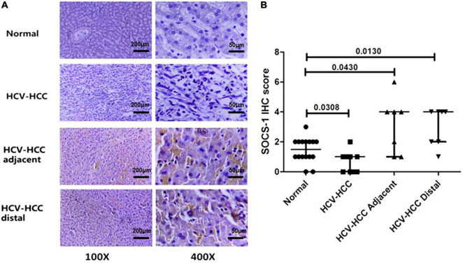
FIGURE 1.
Immunohistochemical staining of suppressor of cytokine signaling 1 (SOCS-1) in different kinds of liver tissues. (A) Representative images of stained SOCS-1 in normal liver tissue, HCV–HCC tissues, HCV–HCC adjacent tissue, and HCV–HCC distal tissue (scale bar = 50 μm in 400× images, 200 μm in 100× images). (B) Axiotis scores of SOCS-1 expression in liver tissues. The enrolled tissues included 10 HCV-HCC tissues, seven adjacent tissues, and seven distal tissues. Also, 16 normal liver tissue sections were obtained as control (Mann–Whitney U test).

