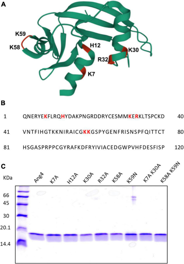
FIGURE 1.

Structural overview and generation of WT and Mutant Angiogenin 4. (A) 3D structure of Ang4 (PDB entry 2J4T). The mutated residues in this study are highlighted in color and labeled. (B) Amino acid sequence of Ang4. (C) SDS-PAGE of purified WT Ang4 and its mutants. Proteins were electrophoresed on a 15% polyacrylamide gel and stained with the Coomassie Brilliant Blue R-250 dye.
