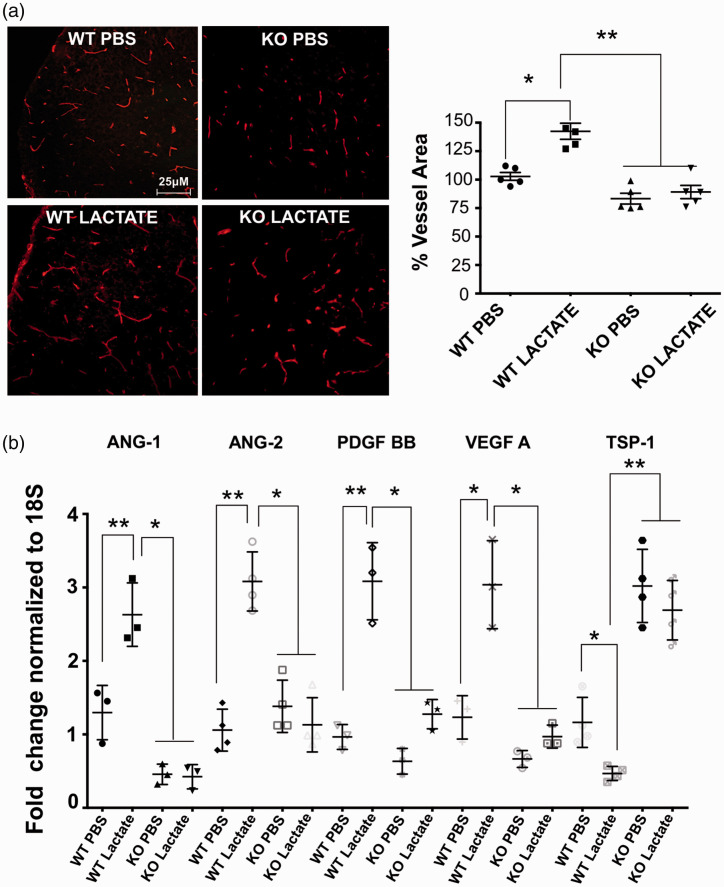
Figure 6.
Lactate acting via GPR81 augments brain vascular density by increasing VEGF expression and suppressing TSP-1. (a) Representative confocal images of lectin-stained (red) brain cross-sections (in P5) showing a marked (25%) increase in cerebral vasculature in the cortex from WT mice compared to age matched GPR81-null mice, 24 hours after intra-cerebroventricular lactate injections. Histogram represent the quantification of the brain vascular area in the cortex area from WT versus KO GPR81-null mice at P5. Values represent mean ± SD; n = 5 animals per group. *p < 0.05, **p < 0.01 compared to WT PBS, one-way ANOVA followed by the Dunnett’s test for multiple comparison with control. (b) Real-time quantitative PCR analysis showing the mRNA expression of proangiogenic factors VEGF, ANG-2, PDGF; anti-angiogenic factor TSP-I and pro-inflammatory factors COX-2 and CCL-2 in WT mice and GPR81-null mice, 24 hours after intra-cerebroventricular lactate injections. Values represent mean ± SD; n = 4–5 samples per group. *p < 0.05, **p < 0.01 compared to their respective control; one-way ANOVA followed by the Dunnett’s test for multiple comparison with control.

