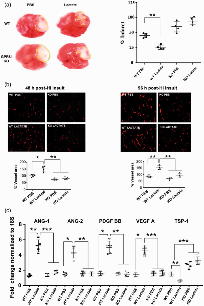
Figure 7.
Lactate reduces infarct size and concordantly augments cerebral vascular density following perinatal HI insult. (a) Tetrazolium chloride (TTC) staining in brains showing infarct size (dotted lines) in WT compared to GPR81-null mice after intra-cerebroventricular lactate (10 mmol/L) injections. Histograms represent infarct size (percentage) in WT versus GPR81-null mice. Values represent mean ± SD; n = 4 animals per group. **p < 0.01 compared to WT PBS; one-way ANOVA followed by the Dunnett’s test for multiple comparison with control. (b) Representative confocal images of lectin-stained (red) brain cross-sections (in P5) showing vasculature mostly in the periinfarct area of cortex from WT mice and age matched GPR81-null mice pre-treated with lactate or PBS (vehicle) and evaluated after 48 hours and 96 hours post-HI. Histograms represent percentage of vessel area in WT versus GPR81-null mice after lactate treatment. Values represent mean ± SD; n = 4 animals per group. *p < 0.05, **p < 0.01 compared to WT PBS; one-way ANOVA followed by the Dunnett’s test for multiple comparison with control. (c) Real-time quantitative PCR analysis showing the mRNA expression of proangiogenic factors VEGF, ANG-1, ANG-2, PDGF; anti-angiogenic factor TSP-I in brain tissues from WT and GPR81-null mice, 48 hours after HI insult and intra-cerebroventricularly injected or not with lactate. Data represent mean ± SD; n = 5 samples per group; *p < 0.05, **p < 0.01, ***p < 0.001 compared with WT PBS, one-way ANOVA followed by the Dunnett’s test for multiple comparison with control.

