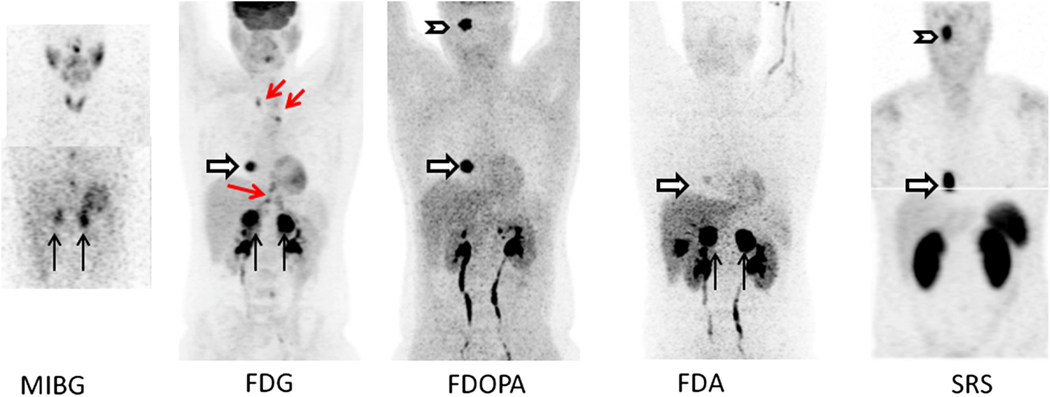
Fig. 2.
Patient with SDHD and metastatic PHEO/PGL. Imaging with a variety of radiopharmaceutical shows heterogeneity in uptake. MIBG panel (MIP—top, and coronal slice—bottom) shows uptake in bilateral adrenal PHEOs (black arrows). Two foci in lower midline chest are thought to be due to swallowed activity in esophagus, glomus tumor is not visualized. FDG MIP panel shows intense bilateral uptake in the adrenal PHEOs and a right atrial PGL (white arrow). Mild uptake in a right glomus jugulare PGL was also seen (not shown this projection). Foci in mediastinum and upper abdomen (red arrows) represent uptake in brown fat. FDOPA MIP panel shows intense uptake in right glomus jugulare PGL (chevron) and right atrial lesion. Mild uptake was seen in the left adrenal PHEO but not right (not seen in this view). FDA MIP shows intense uptake in bilateral adrenal PHEOs with faint uptake in atrial lesion. Physiologic gallbladder activity is also seen. No intracranial uptake was seen. SRS panel shows In-111 pentetreotide MIP images with intense uptake in the right glomus jugulare tumor and cardiac lesion but no uptake in the bilateral adrenal tumors. Images courtesy of Dr. Karel Pacak, NICHD, NIH.

