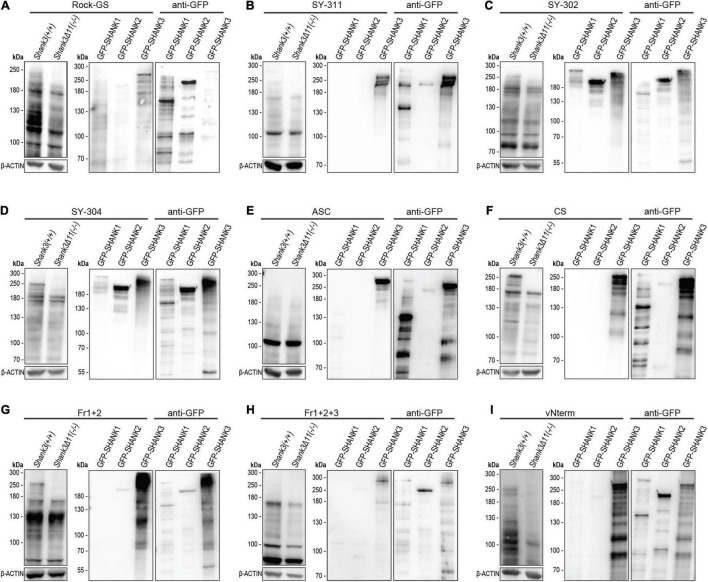
FIGURE 2.
Western Blot analysis for SHANK3 specificity. (A) Rock-GS, (B) SY-311, (C) SY-302, (D) SY-304, (E) ASC, (F) CS, (G) Fr1+2, (H) Fr1+2+3, and (I) vNterm. Left: The Western Blot shows mouse cortical lysate of WT and KO mice for all antibodies. Same amounts of protein are ensured by β-ACTIN. Right: GFP-SHANK1, GFP-SHANK2, and GFP-SHANK3 were overexpressed in HEK293T cells. Blot were first incubated with the respective SHANK3 antibody and then with an anti-GFP antibody to ensure plasmid expression.

