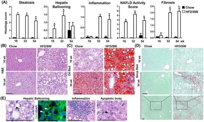
FIGURE 2.

Chronic HFD/SW feeding leads to liver steatosis, steatohepatitis, and fibrosis. (A) Histology scores for steatosis, hepatic ballooning, inflammation, fibrosis, and NAFLD activity score at 16, 32, and 54 weeks for mice fed chow diet (n = 3) or HFD/SW (n = 7–10). Data are expressed as means ± SEM. *p < .05 compared to the chow diet at the respective timepoints. (B–D) Representative liver sections stained with H&E, Oil‐Red‐O, and Sirius Red, respectively. (E) Representative H&E images of hepatic ballooning, inflammation, and apoptotic bodies. Hepatic ballooning is also depicted as loss of cytoplasmic CK18 staining (white arrow). Scale bar: 100 µm. CK18, cytokeratin 18; NAFLD, non‐alcoholic fatty liver disease; SW, sugar water.
