
FIGURE 4.
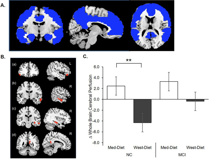
Diet effects on cerebral perfusion assessed with pseudo‐continuous arterial spin labeling (pcASL) magnetic resonance imaging (MRI). A, Rendering of the a priori anatomical mask used in the study constructed using WFU PickAtlas toolbox. 30 The mask included the bilateral superior, inferior, middle and medial frontal cortices; superior, middle, and inferior temporal gyrus; posterior cingulate, precuneus, parahippocampal gyrus; amygdala; and hippocampus. B, The Mediterranean diet (Med‐diet) was associated with significantly greater cerebral perfusion following diet intervention compared to the Western diet (West‐diet) group in the (a) left inferior frontal cortex and the right temporal lobe (b), hippocampus (c), and precuneus (d). C, Normal cognition (NC) participants showed increased mean cerebral perfusion after the Med‐diet, and decreased perfusion after the West‐diet (P = .003). Although the mild cognitive impairment (MCI) group perfusion values also increased following the Med‐diet, this effect was variable and did not approach significance (P = .499). Figures depict change scores for ease of interpretation, adjusted means from repeated measures analysis of covariance are included in Table S2. Error bars represent ± 1 standard error from the mean. Significance is set at + P < .10, *P < .05, ** P < .01, or *** P < .001