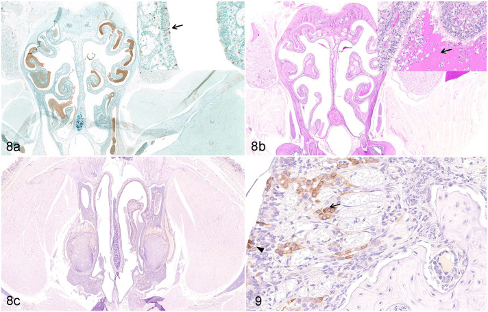
Figures 8 and 9.
SARS-CoV-2 infection, nasal mucosa, Syrian hamster, 7 days post-infection. (a) Apoptosis is detected in the nuclear layer of sustentacular cells (arrow, inset) in the olfactory epithelium. TUNEL. (b) A large amount of mucus is accumulated on the surface of the olfactory epithelium in the nasal cavity (arrow, inset). Periodic acid-Schiff. (c) and Figure 9. F4/80-positive macrophages are detected in mucous layer (black arrowhead, Fig. 9) and submucosa (arrow, Fig. 9) of the necrotic olfactory epithelium. Immunohistochemistry (IHC) for F4/80.
Abbreviations: SARS-CoV-2, severe acute respiratory syndrome coronavirus-2.

