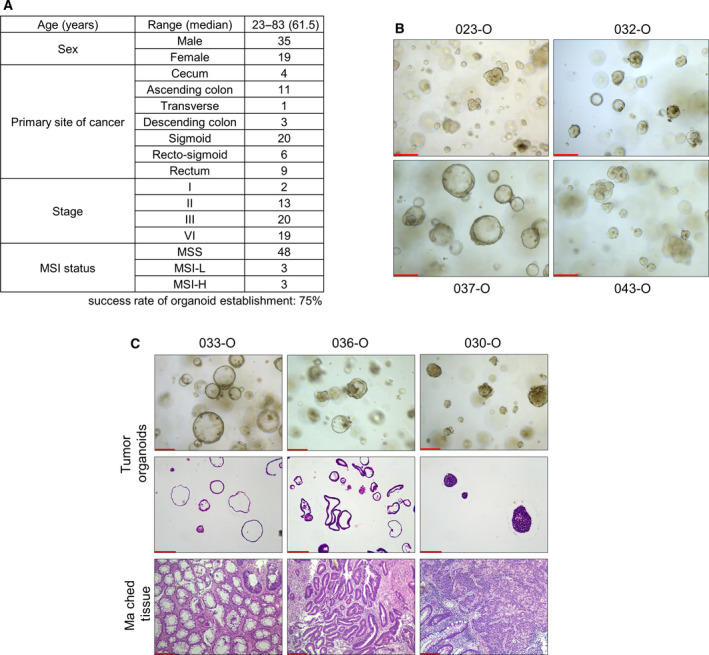
Fig. 1.

Establishment of organoids from patients with colorectal cancer. (A) Summary of patient information in this tumor organoid study. Organoids were derived from variable colon and rectum location with 75% of success rate. (B) Representative images of tumor organoid morphology derived from different patients. PDOs exhibited varying morphology, namely a cystic (e.g., 037‐O, n = 3), aggregated (e.g., 043‐O, n = 3), or mixed form (e.g., 032‐O, n = 4). Scale bar = 200 µm. (C) Representative images of PDOs compared with H&E‐stained matched original tumor tissues and organoids (n = 2). The cultured PDOs and matched tissues showed similar morphology. Scale bar = 200 µm. H&E, hematoxylin and eosin; MSI‐H, microsatellite instability‐high; MSI‐L, microsatellite instability‐low; MSS, microsatellite stability; PDOs, patient‐derived organoids.
