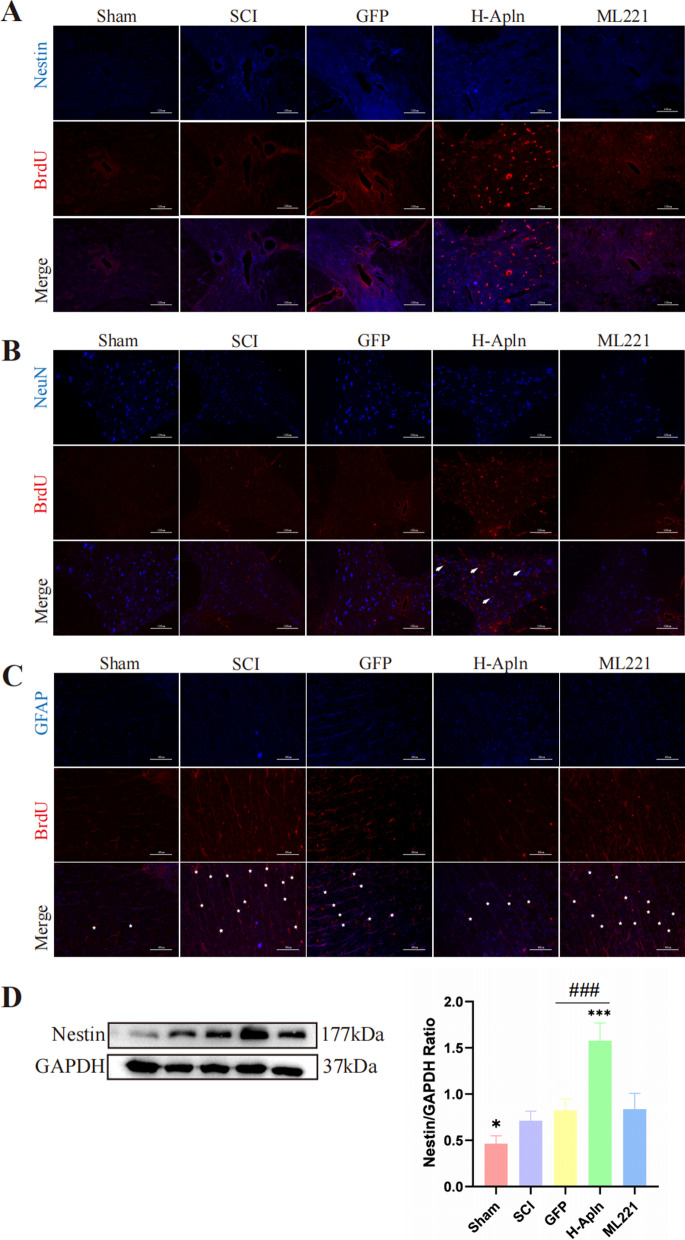
Fig. 8.
Activation and differentiation of endogenous neural stem cells after transplantation of iPSCs. A Activation and proliferation of endogenous NSCs was detected using BrdU (red)/Nestin (blue) co-immunofluorescence at 14 days after SCI in each group. B, C Double-immunofluorescence staining of NeuN (red) and BrdU to identify the differentiation of newly formed cells towards neurons (arrowheads), or GFAP (blue) and BrdU (red) to identify newly formed astrocytes (asterisk). D Western blotting analysis of Nestin protein expression (*p < 0.05 vs. SCI group, ###p < 0.001, GFP vs. H-Apln, mean ± SD)

