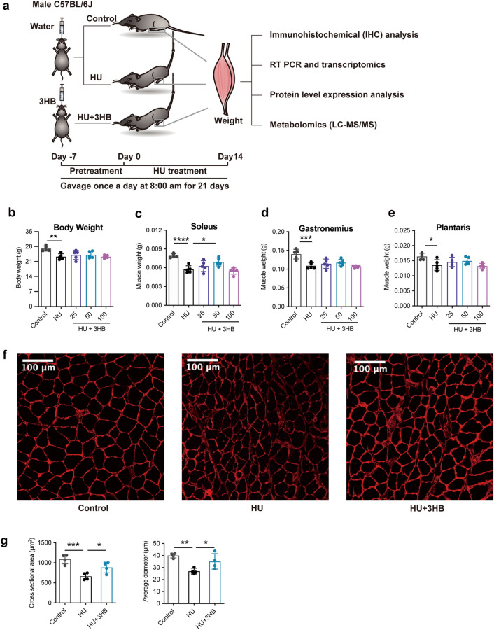
Fig. 1.
3HB preserves soleus muscle mass in hindlimb unloading mice. a Schematic diagram of the development of the hindlimb unloading mouse model. C57BL/6J male mice (8 weeks) were gavage fed with (R)-3-hydroxybutyrate (3HB) 25, 50, and 100 mg/kg body weight once a day for 21 days including 7 days for 3HB pretreatment and 14 days during the hindlimb unloading process. control (ground control group without hindlimbs suspended), HU (hindlimb unloading group), and HU + 3HB group (hindlimb unloading mice fed with 50 mg/kg/days 3HB). Muscle tissues were sampled later for subsequent analyses. b Body weight and muscle mass of c Soleus, d Plantaris, and e Gastrocnemius after 14 days of HU treatments (n = 5). f Images of immunohistochemical (IHC) analysis in laminin-stained muscles (n = 4 per group). Scale bars = 100 µm. g Statistical results of the mean cross-sectional area (CSA) and mean soleus fiber area (n = 4). Error bars are represented as mean ± SD. One-way ANOVA was used for comparison between groups. ****P < 0.0001, ***P < 0.001, **P < 0.01, *P < 0.05, compared with HU mouse group

