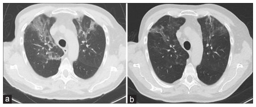
Figure 1:

Imaging of PJP infection. Computed tomography scan of the thorax showing diffuse interstitial infiltrates bilaterally correlating with (a) Pneumocystis jiroveci pneumonia and (b) clearing of these infiltrates after 2 weeks of trimethoprim/sulfamethoxazole. PJP: Pneumocystis jiroveci pneumonia.
