
Figure 3.
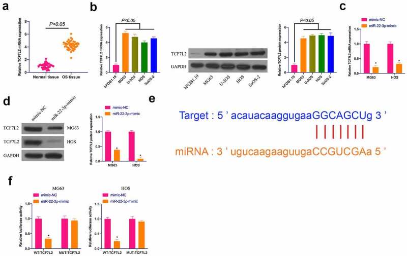
TCF7L2 is highly expressed in OS and targeted by miR-22-3p. A. TCF7L2 expression in OS tissues and adjacent normal tissues detected by RT-qPCR; B. TCF7L2 in OS cell lines MG63, U-2OS, HOS and SAOS-2 detected by RT-qPCR and Western blot; C, D. TCF7L2 in MG63 and HOS cells after transfection of miR-22-3p mimics detected by RT-qPCR and Western blot; E. MiR-22-3p and TCF7L2 potential binding sites queried through bioinformatics website http://starbase.sysu.edu.cn/; F. Targeting relationship between miR-22-3p and TCF7L2 detected by dual luciferase assay; The values were shown as mean ± SD (n = 3). The significance of each group was calculated using one-way ANOVA, and the variance correction via Tukey’s test. Vs. the mimic-NC group, * P < 0.05.