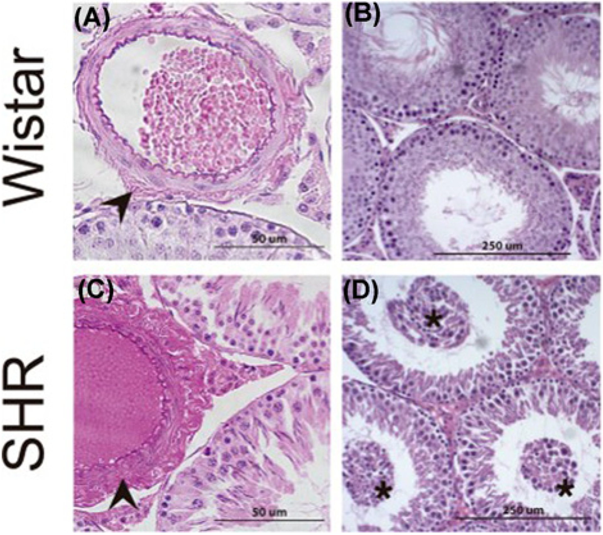
Figure 2. Hematoxylin and eosin staining of testis from normal and spontaneously hypertensive rats.

(A) Normal histoarchitecture of testes from 26-week-old Wistar rats. Arrowheads indicate arteriolar adventitia. (B) Testes from 26-week-old Wistar rats showing intact seminiferous tubules and interstitial compartment. (C) A significant increase in adventitial layers in testicular arterioles (arrowheads) independent of their diameter in spontaneously hypertensive rats (SHR). (D) Testes from SHR showing immature germ cells in the tubular lumen (asterisk). The images are reproduced from the reference [51] with permission.
