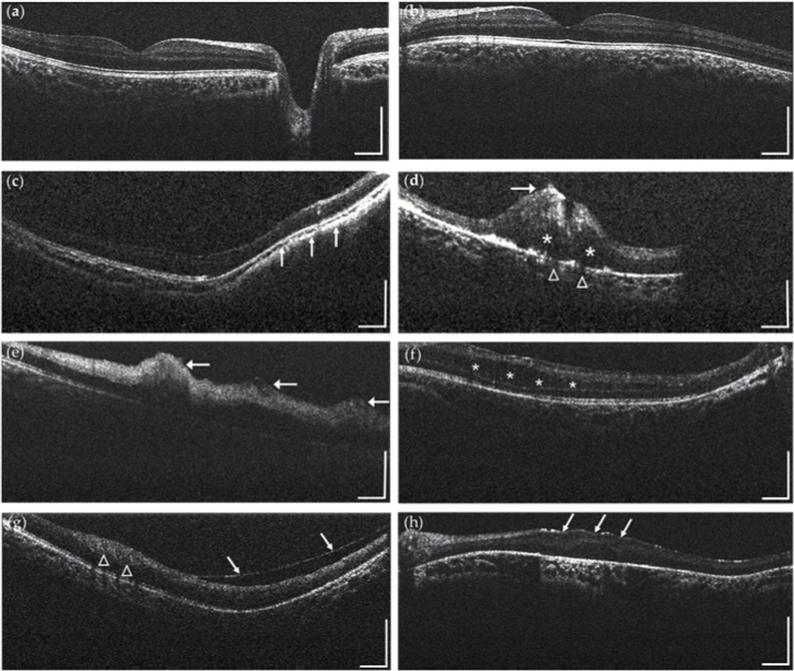
Fig. 6.
Representative structural OCT imaging results obtained from pediatric patients: (a-b) healthy infant; (c) high myopia patient with retina deformation and choroidal thinning (arrow); (d) viral retinitis patient with intra-retinal oedema (asterisk), disruptive inner retina layers(arrow) and choriocapillaris alteration (triangle); (e) retinal hemorrhages patient with hemorrhages appearing hyper reflective and present in multiple retinal layers(arrow); (f) FEVR patient with intra-retinal separation (asterisk) along the outer nuclear layer of the retina. (g) ERM patient with local thickening of the retina (triangle), and epiretinal membrane separate from the inner retina (arrows); (h) LCA patient with upward deformation and disorganization of the retina in the macular area (arrows). All scale bars: 1 mm.

