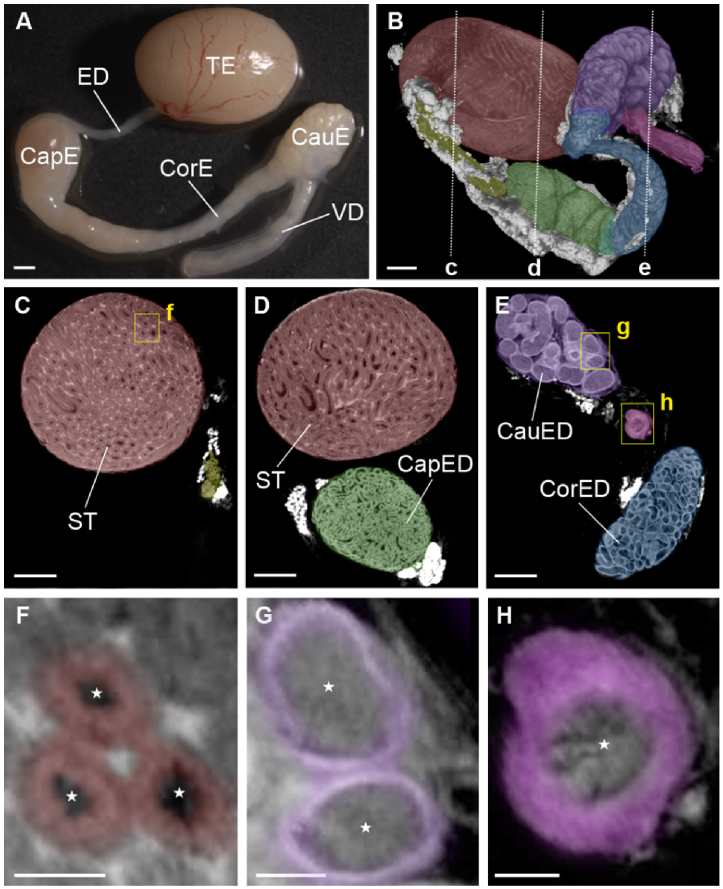
Fig. 1.
Overall structure of the mouse male reproductive tract. (A) Bright-field microscopy image of the male reproductive tract, including the testis (TE), efferent ducts (ED), caput epididymis (CapE), corpus epididymis (CorE), cauda epididymis (CauE), and vas deferens (VD). (B) Micro CT imaging of the male reproductive tract. The dashed line represents the location for the corresponding cross-section shown in panels C-E. Different organs and distinct regions of the epididymis are color-coded: testis (red), efferent ducts (yellow), caput (green), corpus (blue), cauda (purple), and vas deferens (magenta). (C-E) Corresponding depth-resolved micro CT images of the male reproductive tract revealing microstructural details such as seminiferous tubules (ST), caput epididymal ducts (CapED), corpus epididymal ducts (CorED), and cauda epididymal ducts (CauED). The yellow rectangles represent the location for the corresponding magnified images shown in panels F-H. Corresponding magnified images of seminiferous tubules (F), cauda epididymal ducts (G), and the duct of vas deferens (H). The color coding correspond to germ cells (red), epididymal epithelium (purple), and muscle layers of vas deferens (magenta). The stars in (F-H) indicate the lumens of the seminiferous tubules, epididymal ducts, or vas deferens, respectively. Scale bars in (A,B) correspond to 1000 µm, in (C-E) to 500 µm, and in (F-H) to 200 µm.

