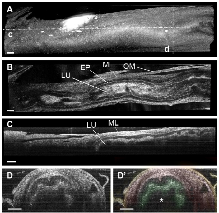
Fig. 4.
3D reconstructions of mouse vas deferens. (A) Three-dimensional OCT image of the vas deferens showing the external morphology. The dashed lines represent the location for the cross-section shown in panels C-D. (B) OCT cross-sectional image of the vas deferens showing the lumen (LU), epithelium (EP), smooth muscle layers (ML), and the outer membrane (OM) of the duct. (C) Corresponding longitudinal OCT image of the vas deferens. (D) Corresponding depth-resolved cross-sectional images of the mouse vas deferens. Different structural features were color-coded: epithelium (green), muscle layers (red), and outer membrane (yellow). The star indicates the inner lumen of the duct. All scale bars correspond to 200 µm.

