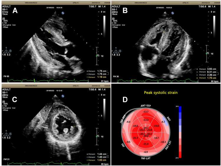
Figure 3.
(A) Subcostal long-axis view showed the anterior ventricular septum and posterior wall were significantly thickened and a characteristic “granular sparkling” appearance of the thickened cardiac walls. (B) Subcostal 4-chamber view showed the ventricular septum and posterior lateral wall were significantly thickened and a characteristic “granular sparkling” appearance of the thickened cardiac walls. (C) Subcostal short axis view at the level of papillary muscle: all of the basal segments of ventricular wall were significantly thickened with a characteristic “granular sparkling” appearance. (D) The Bull’s eye diagram showed reduced global longitudinal strain (GLS) with apical sparing.

