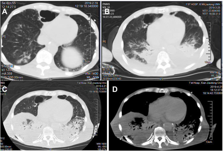
Figure 4.
Display of chest CT images of the patient. Figure (A–C) were displayed on lung window at the top of the diaphragm at different times. The small calcification shadow on the lung surface at the base of both lungs gradually increased. In additional, they all showed diffuse pulmonary interstitial changes, cystic change and lung consolidation shadows. (D) Mediastinal window on the same plane as figure (A–C): fine sand granular calcification can be seen under the pleura in the lesion of the lower lobe of the left lung, which is clearer than that in the lung window.

