. 2022 Jun 20;11:e76027. doi: 10.7554/eLife.76027

Figure 4. Adhesive strength between the biofilm and the substrate governs channel formation.

(a) Reconstructions from laser-scanning confocal microscopy images of the biofilm. The biofilms are either grown on a low surface free energy PDMS substrate (left, γPDMS=23mNm-1) or on a high surface free energy PDMS substrate (right, γPDMS=37mNm-1). (b) Average effective biofilm thickness as a function of time and surface free energy. The average thickness of biofilm grown on a low surface free energy PDMS substrate (red) and on a high surface free energy PDMS substrate (blue). (c) The image sequence shows a biofilm that is grown on a patterned PDMS substrate in the same microfluidic channel. On the left, the substrate has a low surface free energy, while on the right it has a high surface free energy.

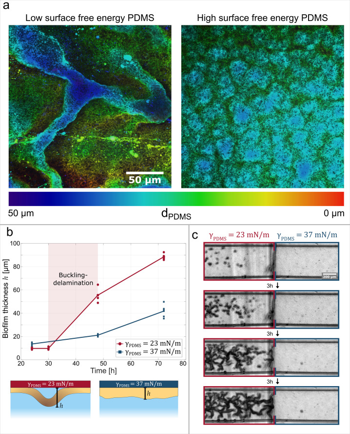
Figure 4.

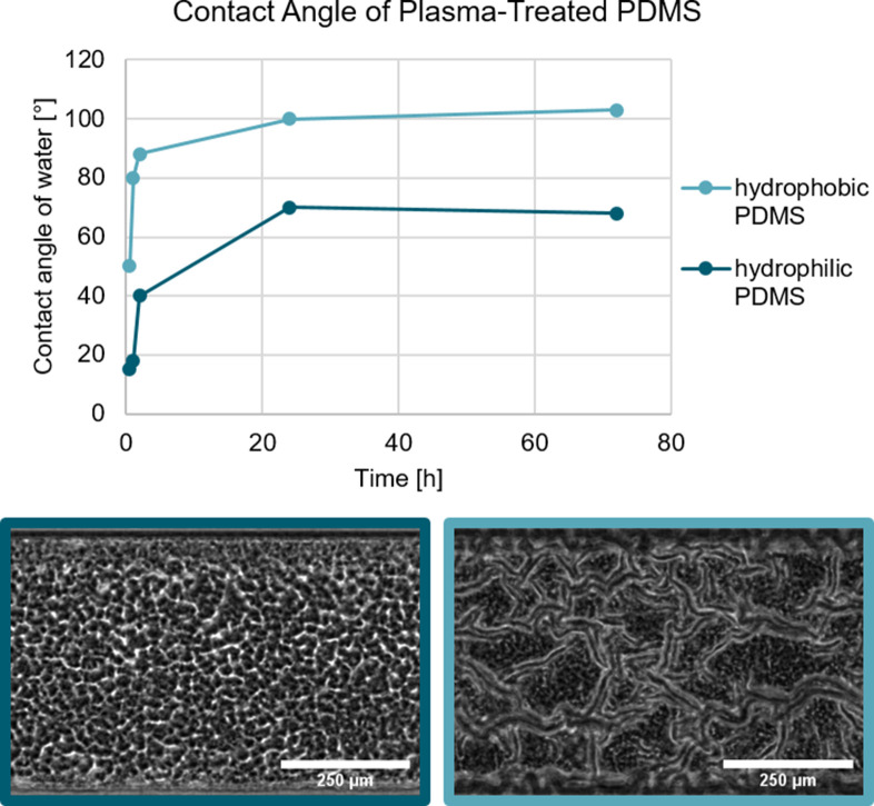
Figure 4—figure supplement 1. The surface free energy of the substrate can be changed through a physical process.

Figure 4—figure supplement 1.

The treatment of PDMS with an oxygen plasma increases the hydrophilicity with identical results to the chemical modification. Biofilm growth on hydrophilic and hydrophobic PDMS substrate. In both cases, the PDMS was treated with an oxygen plasma which renders the PDMS hydrophilic. As shown by Kim et al., 2004 the hydrophobic recovery of plasma-treated can be varied depending on the storage conditions. Biofilm grown on the hydrophilic PDMS does not form a channel network, while the biofilm grown on the hydrophobic PDMS does form a channel network.