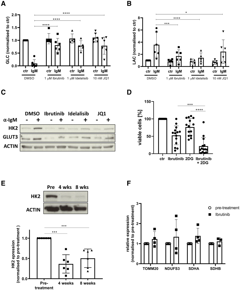
Figure 4.
Inhibition of BCR-signaling and MYC transcription metabolically reprogram CLL cells in vitro and in vivo. (A, B) CLL cells (n = 6) were cultured for 48 hours in the presence of the inhibitors or vehicle control (DMSO) and the residual concentration of glucose (A) and lactate (B) in the media was assessed. There was a significant reduction in glucose and increase in lactate with anti-IgM stimulation, which was inhibited by ibrutinib, idelalisib, and JQ1. (C) Representative blot demonstrating the impact of the inhibitors on the induction of HK2 and GLUT3 after anti-IgM stimulation for 24 hours. The induction of HK2 (n = 7) and GLUT3 (n = 7) was also inhibited by ibrutinib, idelalisib, and JQ1. (D) CLL cells (n = 14) were cultured for 48 hours in the presence of 10 µM ibrutinib (or vehicle control—DMSO) and 1 mM 2-deoxyglucose (2-DG) as indicated and apoptosis was assessed by Annexin V binding. Addition of 2-DG resulted in increased sensitivity to ibrutinib. (E) The impact of ibrutinib on the expression of HK2 in peripheral blood CLL cells was assessed in previously untreated patients initiating treatment with single-agent ibrutinib. Representative blot of HK2 showing a reduction in expression at 4 and 8 weeks of treatment in vivo. The expression of HK2 decreased significantly in all cases after 4 weeks (n = 6) and 8 weeks (n = 6) of treatment. (F) In contrast, the expression of TOMM20 (n = 4), NDUFS3 (n = 4), SDHA (n = 4), and SDHB (n = 3) did not change after ibrutinib treatment. BCR = B-cell receptor; CLL = chronic lymphocytic leukemia; DMSO = dimethyl sulfoxide; SDHA, succinate dehydrogenase complex subunit A; SDHB, succinate dehydrogenase complex subunit B.

