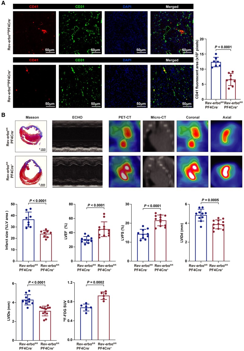
Figure 5.
Platelet-specific deletion of Rev-erbα protected against microvascular microthrombi obstruction and infarct expansion. (A) Immunofluorescence of the hearts from Rev-erbαfl/flPF4Cre− and Rev-erbαfl/flPF4Cre+ post-acute myocardial infarction. Platelets, endothelial cells and nuclei were stained with anti-CD41, anti-CD31, and 4',6-diamidino-2-phenylindole. Scale bars = 50 μm. Quantification of CD41 area per field (n = 7–8 per group). Data were analysed by Student's t-test. (B) Representative images of Masson's trichrome-stained hearts, M-mode echocardiography, and positron emission tomography computed tomography scanning of Rev-erbαfl/flPF4Cre− and Rev-erbαfl/flPF4Cre+ mice on day seven post-acute myocardial infarction. Quantification of infarct size (n = 8 per group), echocardiographic cardiac function (n = 10–12 per group), and mean myocardial 18F-FDG SUV (n = 6 per group). Data were analysed by Student's t-test. ECHO indicates echocardiography; 18F-FDG SUV, 18F-fluorodeoxyglucose standardized uptake value; LV, left ventricular; LVEF, left ventricular ejection fraction; LVFS, left ventricularfractional shortening; LVIDs, left ventricularinternal dimension in systole; LVIDd, left ventricular internal dimension in diastole; PET-CT, positron emission tomography computed tomography.

