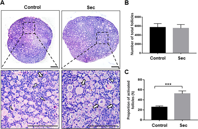
Fig. 1.
The hUC-MSC-sec promoted primordial follicle activation in vitro. A. After 12 days of in vitro culture, the histological analysis of hematoxylin and eosin staining showed more activated follicles in hUC-MSC-sec-treated ovaries than controls. The arrowheads indicate primordial follicles, and the arrows indicate activated follicles. B. Quantification of ovarian follicles showed no obvious difference in the total number of follicles between hUC-MSC-sec-treated ovaries (5510 ± 832.2) and controls (5723 ± 825.3). C. Ovarian follicle counts revealed a significantly increased proportion of activated follicles in hUC-MSC-sec-treated ovaries (52.4 ± 5.3%) compared to controls (25.5 ± 2.3%). Data are shown as the mean ± SD, n = 6. ***P < 0.001. Scale bars, 100 μm

