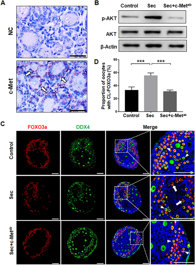
Fig. 4.

The hUC-MSC-sec promoted the activation of the PI3K-AKT pathway through c-Met on the granulosa cells. A. Immunostaining showing that c-Met is expressed on granulosa cells in both primordial and activated follicles. Normal mouse IgG was used in place of the primary antibody for the negative control. B. Western blot showing the expression of p-AKT in mouse ovaries after 4 days of in vitro culture. The ability of the MSC-sec to increase the phosphorylation of AKT was greatly inhibited by the addition of c-Metab. C. Immunofluorescence analysis showing the location of FOXO3a in mouse ovaries after 4 days of in vitro culture. The ability of the hUC-MSC-sec to promote translocation of FOXO3a from the nucleus to the cytoplasm was inhibited by the addition of c-Metab. The arrowheads indicate the nuclear localization of FOXO3a, and the arrows indicate the cytoplasmic localization of FOXO3a. D. The proportion of CL-FOXO3a was significantly decreased in hUC-MSC-sec plus c-Metab-treated ovaries (30.8 ± 2.5%), which was similar to controls (32.7 ± 5.3%), compared to the hUC-MSC-sec group (55.2 ± 4.4%). Data are shown as the mean ± SD, n = 6. ***P < 0.001. Scale bars in A, 20 μm; Scale bars in C, 100 μm
