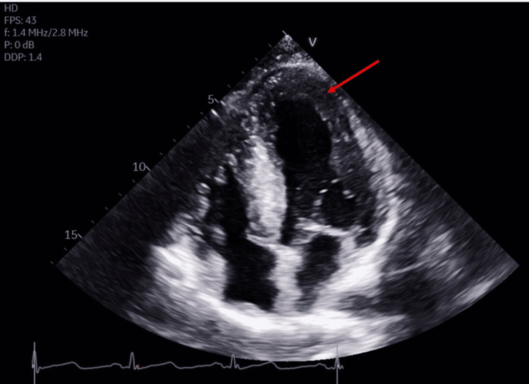
Figure 1. Transthoracic echocardiogram.
Transthoracic echocardiogram (TTE) in apical four-chamber view shows severe concentric left ventricular hypertrophy (LVH) (arrow). The ejection fraction was 60-65%. All segments contract normally. The diastolic filling pattern indicates impaired relaxation and elevated left ventricular end-diastolic pressure.

