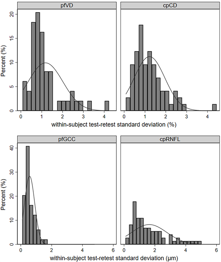
Figure 1.
Distribution of the within-subject test–retest SD for optic nerve head (left) and macula (right), and vessel density (upper) and thickness (lower). cpCD, circumpapillary capillary density; cpRNFL, circumpapillary retinal nerve fibre layer; pfVD, parafoveal vessel density; pfGCC, parafoveal ganglion cell complex.

