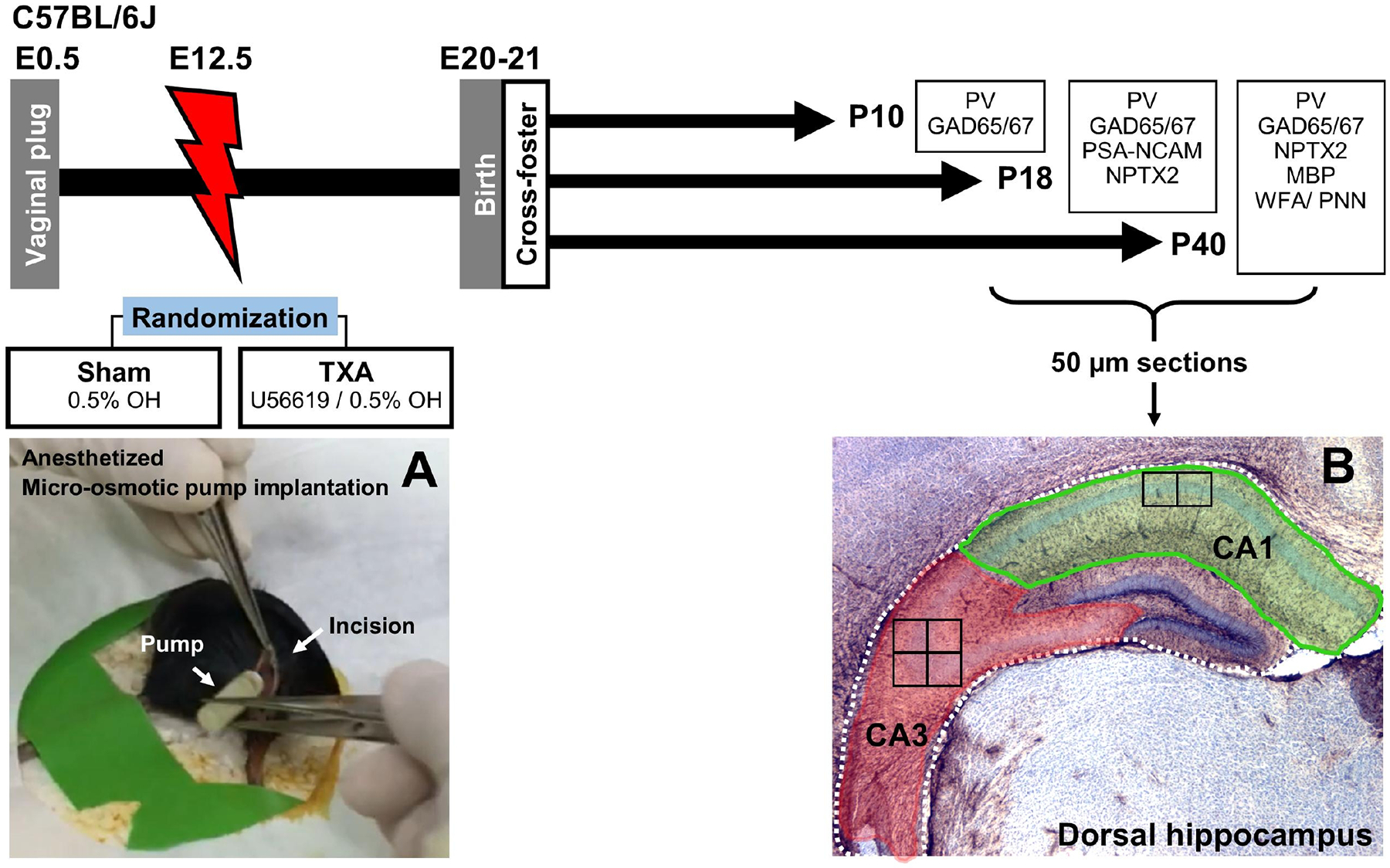
FIGURE 1. Experimental design.

Impregnation of C57BL6/J dams was confirmed by visualizing vaginal plug (E0.5). Under anesthesia micro-osmotic pumps containing either vehicle (0.5% ethanol, OH) were implanted at E12.5 (A). Pups were born at P20–21 and were cross-fostered to unmanipulated dams. For these experiments, 3 groups of mice of both sexes were survived to P10 (n=4 per treatment per sex), P18 (n=7 per treatment per sex), or P40 (n=7 per treatment per sex). Following perfusion, brains were collected, post-fixed, cryoprotected, and frozen. Brains were sectioned coronally in a freezing microtome at 50μm. Anterior sections with embedded dorsal hippocampus were used for experiments evaluating target proteins essential for initiation, consolidation, and closure of the CPd of synaptic plasticity in the CA1 and CA3 subfields (B). Location of the high magnification fields evaluated are depicted as squares inside the CA1 (x2) and CA3 (x4). CA, cornus ammonis; GAD, Glutamic acid decarboxylase; E, embryonic day; MBP, Myelin basic protein; NPTX2, neuronal pentraxin 2; P, post-natal age; PNN, perineural nets; PSA-NCAM, polysialylated neural cell adhesion molecule; PV, parvalbumin; TXA, thromboxane A2-analog; WFA, Wisteria floribunda lectin.
