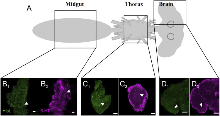
FIGURE 1.
Serotonergic and histaminergic innervation of A. stephensi tissues. (A) Schematic of the regions for immunohistochemistry. (B) Staining of the midgut for histamine (B 1 ) and 5-HT (B 2 ). Arrows denote the differences in staining for the lining of the midgut. (C) Staining of the thoracic ganglion for histamine (C 1 ) and 5-HT (C 2 ). Arrows denote blebby-like staining. (D) Staining of the brain for histamine (D 1 ) and 5-HT (D 2 ). Arrows denote stronger staining in the optic lobes for histamine and 5-HT.

