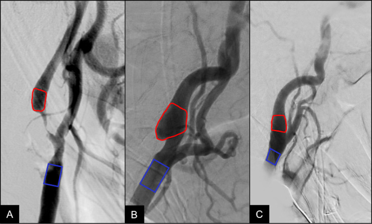
Figure 1.
Region of interest placement: representative lateral digital subtraction angiography image of a patient with severe carotid atherosclerosis (A) and carotid web (B) with color-coded red region of interest (ROI) distal to the lesion and a second blue ROI placed at the distal common carotid artery bifurcation for calculation of normalized hemodynamic parameters. A normal carotid bifurcation (C) and ROI placement is shown for comparison.

