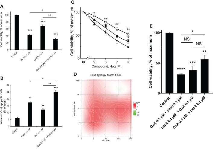
Figure 2.
Inhibition of AsPC-1 cell growth by OxA and/or Nab-paclitaxel. (A) Inhibition of AsPC-1 cell viability induced by 0.1 µM Nab-paclitaxel (pacli), 0.1 µM OxA (OxA), or 0.1 µM Nab-paclitaxel plus 0.1 µM OxA. Results were expressed in the percentage of maximum obtained in the absence of treatment (control). (B) Effect of OxA or Nab-paclitaxel or their addition on apoptosis of ASPC-1 cells. Apoptosis was determined by annexin V/7-AAD binding, and results were expressed as the percentage of apoptotic cells. (C) Effect of the dose–response of OxA or Nab-paclitaxel or OxA plus Nab-paclitaxel on cell viability of AsPC-1 cells. (D) Bliss model diagram and estimation of Bliss synergy score. (E) OxA plus Nab-paclitaxel treatment and Impact of sequential treatment by OxA>Nab-paclitaxel or Nab-paclitaxel>OxA compared to control (absence of treatment). Results were expressed in the percentage of maximum obtained in the absence of treatment. Data were the means ± SEM of three separate experiments. (○) OxA, (●) Nab-paclitaxel, and (▲) OxA plus Nab-paclitaxel. NS, non-significant, *p<0.05, **p<0.01, ***p<0.001, and ****p<0.0001 corresponding to comparison with control.

