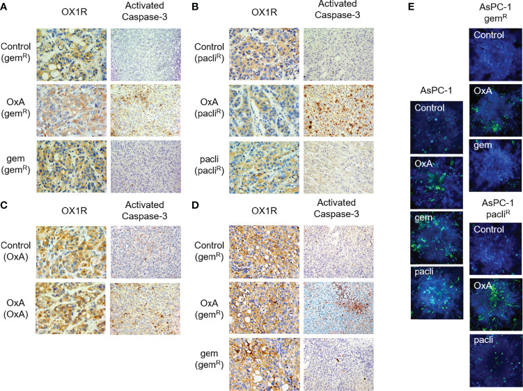
Figure 6.
Histological analysis of resected tumors from Run 02 experiments. (A) OX1R expression and caspase-3 activation in gemcitabine-chemoresistant AsPC-1 tumors (gemR) treated with PBS (Control), OxA (OxA), and gemcitabine (gem). (B) OX1R expression and caspase-3 activation in Nab-paclitaxel-chemoresistant AsPC-1 tumors (pacliR) treated with PBS (Control), OxA (OxA), and Nab-paclitaxel (pacli). (C) OX1R expression and caspase-3 activation in 50 days OxA-treated tumors (OxA) treated with PBS (control) and OxA (OxA). (D) OX1R expression and caspase-3 activation in gemcitabine-chemoresistant tumors obtained by xenografting of 50 days gemcitabine-treated PDAC15 cells (gemR) treated with PBS (control), OxA (OxA), and gemcitabine (gem). (E) Cell viability determination of spheroids developed with parental AsPC-1, gemcitabine-resistant, and Nab-paclitaxel-resistant AsPC-1 cells isolated in Run 02 experiment. Magnification was 40× (OX1R expression) and 20× (caspase-3 activation).

